Custom Search
|
|
 
|
||
|
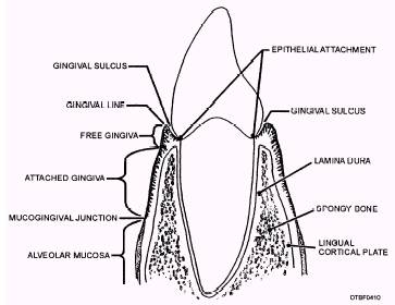
GINGIVA.\The gingiva, shown in figure 4-10, is specialized masticatory mucosa covering the alveolar process. In a healthy mouth, gingiva is firmly in place encircling the necks of the teeth. It aids in the support of the teeth, and protects the alveolar process and periodontal ligament from bacterial invasion. Healthy gingiva is firm and resilient. Healthy gingiva under normal flossing and brushing activities does not bleed. The color of healthy gingiva can range from pale pink to darker shades (purple to black) depending on each individual's pigmentation. The surface of the attached gingiva and interdental papillae may be stippled (resembling the texture of the skin of an orange). Like the tongue, the gingiva is highly vascular and receives its blood supply from the lingual, mental, buccal, and palatine arteries. Other important aspects of the gingiva are discussed in the following paragraphs. Unattached Gingiva.\The portion of gingiva that extends from the gingival crest to the crest of the bone is called unattached gingiva. It can also be called the free gingiva. It can be displaced and is not bound directly to the tooth or bone. In a healthy mouth, this
Figure 4-10.\Structures of the gingiva. portion is approximately 1 to 3 mm wide and forms the soft tissue wall of the gingival sulcus next to the tooth. Other structures of unattached gingiva include: Gingival margin\The 1 mm narrow band of gingiva that forms the immediate collar around the base of the tooth. This area is first to show symptoms of gingivitis. Gingival sulcus\Area between the unattached gingiva and the tooth. Popcorn hulls get trapped in this area. Epithelial attachment\Joins the gingiva to the tooth surface. Interdental papilla\The portion of the free gingiva that fills the interproximal embrasures below the contact areas of adjacent teeth. It helps prevent food from packing between the teeth. Attached Gingiva.\Located apical to the free gingiva on the labial and lingual aspects. It is firmly fixed to the underlying bone of the cortical plates of the alveolar process. Mucogingival Junction.-A line that separates the attached gingiva from the lining mucosa. Lining Mucosa Lining mucosa is found on the inside of the lips, cheeks, vestibule, soft palate, and under the tongue. It consists of a thin, fragile tissue that is very vascular. Lining mucosa is brighter red in color than masticatory mucosa. Also included in the lining mucosa is alveolar mucosa. It lies apical to the mucogingival junction and is loosely attached. TOOTH MORPHOLOGY This section describes the external features of the teeth. A tooth is defined as "one of the hard, bony appendages that are borne on the jaws...and serve for the seizing and mastication of food, as weapons of offense and defense, etc." In man and the lower animals, the design of the teeth are a reflection of eating habits. Animals, classified according to their eating habits, are carnivorous (flesh eating), herbivorous (plant eating), or omnivorous (eating everything; both flesh and plant). |
||
|
||