Custom Search
|
|
 
|
||
|
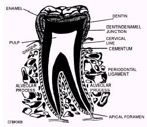
ALVEOLAR PROCESS The alveolar process (fig. 4-8) is that bony portion of the maxilla and mandible where the teeth are embedded and by which tooth roots are supported. The alveolar socket is the cavity within the alveolar process in which the root of the tooth is held by the periodontal ligament. The bone that divides one socket from another is called the interdental septum. When multirooted teeth are present, the bone is called the interradicular septum. The alveolar process includes the cortical plate, alveolar crest, trabecular bone, and the alveolar bone proper. Cortical Plate Structurally, the cortical plate is composed of lingual and facial plates of compact bone. It is dense in nature and provides strength and protection and acts as the attachment for skeletal muscles. The mandibular cortical plate is more dense than the maxilla cortical plate and has fewer perforations for the passage of nerves and blood vessels. Alveolar Crest The alveolar crest is the highest point of the alveolar ridge and joins the facial and lingual cortical plates. Trabecular Bone Trabecular or spongy bone lies within the central portion of the alveolar process, and is the less dense,
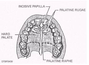
Figure 4-8.\The periodontium. cancellous bone. When viewed by a radiograph, trabecular bone has a web-like appearance. Alveolar Bone Proper The alveolar bone proper is a thin layer of compact bone, that is a specialized continuation of the cortical plate and forms the tooth socket. The lamina dura (fig. 4-10) is a horseshoe shape white line on a dental radiograph that roughly corresponds to the alveolar bone proper. PERIODONTAL LIGAMENT The periodontal ligament (fig. 4-8) is a thin, fibrous ligament that connects the tooth to the bony socket. Normally, teeth do not contact the bone directly; a tooth is suspended in its socket by the fibers of the ligament. This arrangement allows each tooth limited individual movement. The fibers act as shock absorbers to cushion the force of the chewing impact of mastication. TISSUES OF THE ORAL CAVITY The oral cavity is made up of specialized epithelial tissues that surround the teeth and serve as a lining. These tissues are called the oral mucosa and consist of three types: masticatory mucosa, lining mucosa, and specialized mucosa. Masticatory Mucosa Masticatory mucosa is comprised of the tissue that covers the hard palate and the gingiva. Masticatory mucosa is usually light pink in color (can vary with skin color) and is keratinized. Keratinized tissue has a horny, tough, protective outer layer of tissue. Characteristics of masticatory mucosa are: no submucosa lies under the masticatory mucosa, held in place firmly to bone and does not move, has a dense, hard covering, and functions to withstand the active process of chewing and swallowing food. HARD PALATE.\The hard palate or roof of the mouth (fig. 4-9) is covered with masticatory mucosa and is firmly adhered to the palatine process (bone). Its
Figure 4-9.\Masticatory mucosa of the hard palate. color is usually pale pink. Important structures of the hard palate are: Incisive papilla\Located at the midline, directly posterior of the maxillary central incisors (pear-shaped in appearance). Palatine raphe\Extends from the incisive papilla posteriorly at the midline (may be ridge shaped in appearance with a whitish streak at the midline). Palatine rugae\Extends laterally (along side) from the incisive papilla and from the palatine raphe (wrinkled, irregular ridges in appearance). |
||
|
||