Custom Search
|
|
 
|
||
|
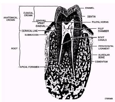
TISSUES OF THE TEETH This section describes the histologic structures of enamel, dentin, cementum, and the dental pulp. Figure 4-6 illustrates the tissues of the teeth. ENAMEL Enamel is translucent and can vary in color from yellowish to grayish white. The different colors of
Figure 4-5.-Bifurcated and trifurcated roots. enamel may be attributed to the variation in the thickness, translucent proprieties, and the quality of the crystal structure and surface stains of enamel. Enamel (fig. 4-6) is the calcified substance that covers the entire anatomic crown of the tooth and protects the dentin. It is the hardest tissue in the human body and consists of approximately 96% inorganic minerals, 1% organic materials, and 3% water. Calcium and phosphorus (as hydroxyapatite) are its main inorganic components. Enamel can endure crushing pressure of approximately 100,000 pounds per square inch. A layering of the dentin and periodontium, coupled with the hardness of the enamel, produces a cushioning effect of the tooth's different structures enabling it to endure the pressures of mastication. Structurally, enamel is composed of millions of enamel rods or prisms. Each rod begins at the dentinoenamel junction (junction between the enamel and dentin) and extends to the outer surface of the crown. Enamel is formed by epithelial cells (ameloblasts) that lose their functional ability when the crown of the tooth has been completed. Therefore, enamel, after formation, has no power of further growth or repair. DENTIN Dentin (fig. 4-6) is the light yellow substance that is more radiolucent than enamel and is very porous; it constitutes the largest portion of the tooth. The pulp chamber is located on the internal surface of the dentin walls. Dentin is harder than bone but softer than enamel. Dentin consists of approximately 70% inorganic matter and 30% organic matter and water. Calcium and phosphorus are its chief inorganic components. Dentin is a living tissue and must be protected during operative or prosthetic procedures from dehydration (drying) and thermal shock. The dentin is perforated by tubules (similar to tiny straws) that run between the cementoenamel junction and the pulp. Cell processes from the pulp reach part way into the tubules like fingers. These cell processes create new dentin and mineralize it. Dentin transmits pain stimuli by the way of dentinal fibers. Because dentin is a living tissue, it has the ability for constant growth and repair that reacts to physiologic (functional) and pathologic (disease) stimuli.
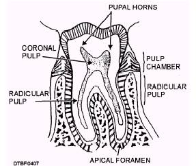
Figure 4-6.\Tissues of the teeth. CEMENTUM Cementum is the bonelike tissue that covers the roots of the teeth in a thin layer (fig. 4-6). It is light yellow in color, slightly lighter than dentin. The cementum is composed of approximately 55% organic material and 45% inorganic material. (The inorganic components are mainly calcium salts.) The cementum joins the enamel at the cervix of the tooth. The point at which they join is called the cementoenamel junction (CEJ). In most teeth the cementum overlaps the enamel for a short distance. In some, the enamel meets the cementum in a sharp line. In a few, a gap may be present between the enamel and the cementum, exposing a narrow area of root dentin. Such areas may be very sensitive to thermal, chemical, or mechanical stimuli. The main function of cementum is to anchor the teeth to the bony walls of the tooth sockets in the periodontium. This is accomplished by means of the fibers of the periodontal ligament or membrane. Cementum is formed continuously throughout the life of the tooth to compensate for the loss of tooth substance because of occlusal wear, and to allow for the attachment of new fibers of the periodontal ligament to the surface of the root. THE DENTAL PULP The dental pulp, (figure 4-7), is the soft tissue of the tooth, which develops from the connective tissue of the dental papilla. Within the crown, the chamber containing the dental pulp is called the pulp chamber. The pulp contains blood vessels and nerves that enter through the apical foramen. The coronal pulp is within the crown. Within the root is the radicular pulp.
Figure 4-7.\Dental pulp. The chief function of the pulp is the formation of dentin. However, it also furnishes nourishment to the dentin; provides sensation to the tooth, and responds to irritation, either by forming reparative secondary dentin or by becoming inflamed. The pulp chamber contains the coronal pulp and pulp horns located within the crown portion of the tooth. The apical foramen is at the end or apex of the radicular pulp. Blood vessels, nerves, and connective tissue pass through this area to reach the interior of the tooth. PERIODONTIUM The tissues that surround and support the teeth are collectively called the periodontium. Their main functions are to support, protect, and provide nourishment to the teeth. Figure 4-8 illustrates the supporting tissues of the periodontium. The periodontium consists of cementum, alveolar process of the maxillae and mandible, periodontal ligament, and gingiva. CEMENTUM Cementum is the only tissue considered as both a basic part of the tooth and a component of the periodontium. It is a thin, calcified layer of tissue that completely covers the dentin of the tooth root. Cementum is forming during the development of the root and throughout the life of the tooth. Cementum functions as an area of attachment for the periodontal ligament fibers. |
 |
|
|
||