Custom Search
|
|
 
|
||
|
Lacrimal Bones The lacrimal bones are the smallest and most fragile of the cranial bones. These thin, scalelike structures are located in back of the frontal process of the maxilla. Nasal Bones The nasal bones are small oblong bones somewhat rectangular in shape. They lie side by side and are fused at the midline to form the bridge of the nose (nasal septum). These bones are responsible for the shape of the nose. Inferior Nasal Conchae The inferior nasal conchae are curved, fragile, scroll-shaped bones that lie in the lateral walls of the nasal cavity. They provide support for mucous membranes within the nasal cavity. Vomer Bone The vomer bone is a thin, flat, single bone almost trapezoid in shape. It connects with the ethmoid bone and together they form the nasal septum. Mandible The mandible (lower jaw-bone) is the longest, strongest, and the only movable bone in the skull. Figure 3-11 illustrates the anatomy of the mandible.
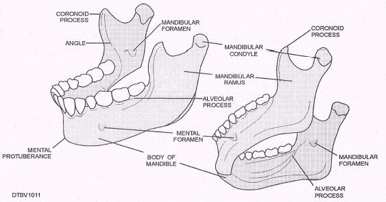
Figure 3-11.\Anatomy of the mandible; lateral view (left), inferior view (right). The mandible is horseshoe-shaped, with an upward sloping portion at each end called the ramus. The rami are divided into two different processes: Condyloid process\Also called mandibular condyle, located posterior on the ramus and forms the head of the mandible. It is knuckle-shaped, and articulates in the glenoid fossa of the temporal bone to form the temporal mandibular joint. Coronoid process\Located anterior of the condyle, and provides attachment for the temporal's muscle, which helps lift the mandible to close the mouth. Other important anatomical landmarks of the mandible you should be able to recognize are as follows: Alveolar process\Supports the teeth of the mandibular arch. Mental protuberance\Also referred to as the chin and is located at the midline of the mandible. Mental foramen\Located on the facial surfaces of the mandible on both the right and left sides, just below the second premolars. This opening contains the mental nerve and blood vessels. Body\The heavy, horizontal portion of the mandible below the mental foramen extending from the angle to the parasyplysis region. Angle\Juncture where the body of the mandible meets with the ramus. Mandibular foramen\Located near the center of each ramus on the medial side (inside), through this opening passes blood vessels and the interior alveolus nerve, which supply the roots of the mandibular teeth. This is a common area where the dental officer will inject anesthetic to block the nerve impulses and make the teeth on that side insensitive (numb). |
 |
|
|
||