Custom Search
|
|
 
|
||
|
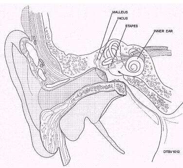
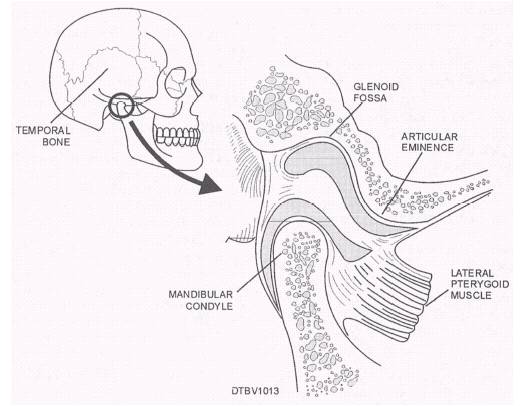
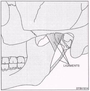
BONES OF THE EAR In each middle' ear and located in the auditory ossicles are three small bones named the malleus, incus, and staples (fig. 3-12). Their function is to transmit and amplify vibrations to the ear drum and inner ear. TEMPORAL MANDIBULAR JOINT The right and left temporal mandibular joints (TMJs) are formed by the articulation of the temporal bone and the mandible. This is where TMJs connect with the rest of the skull. Figure 3-13 illustrates the TMJ. The mandible is joined to the cranium by ligaments of the temporal mandibularjoint (fig. 3-14).
Figure 3-12.\Anatomy of the middle ear.
Figure 3-13.\Temporal mandibular joint.
Figure 3-14.\Ligaments of a temporal mandibular joint. The TMJ consists of three bony parts: Glenoid fossa\Oval depression in the temporal bone that articulates with the mandibular condyle. Articular eminence\Ramp-shaped segment of the temporal bone located anterior to the glenoid fossa. Condyle\The knuckle-shaped portion of the mandibular ramus found on the end of the condyloid process. It is positioned underneath the glenoid fossa and makes up the hinge joint of the TMJ. MUSCLES OF THE HEAD The muscles of the head can be classified into two groups, muscles of facial expression and muscles of mastication. How muscles work and function depends on the action of each muscle (movement), the type of joint it is associated with, and the way the muscle is attached on either side of the joint. Muscles are usually attached to two places: one end being joined to an immovable or fixed portion, and the other end being joined to a movable portion on the other side of a joint. The immovable portion is called the origin of the muscle, and the movable portion is called the insertion. When muscles of the head contract, the insertion end is pulled toward the origin. MUSCLES OF FACIAL EXPRESSION The muscles that are underneath the skin of the face are responsible for helping communicate our feelings through facial expression. The muscles of the mouth help us express surprise, sadness, anger, fear, and pain. Table 3-3 lists the muscles of facial expression and figure 3-15 illustrates these muscles. |
 
|
|
|
||