Custom Search
|
|
 
|
||
|
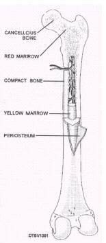
CHAPTER 3 - HEAD AND NECK ANATOMY Basic knowledge of the skull, facial bones, jaws, and muscles of the head and neck region are fundamental for a Dental Technician. It is important to understand the relationship of the bones and muscles as they work together to provide support for the dentition (teeth) and movement for mastication (chewing). STRUCTURE OF BONE The bones of the human skeleton provide rigid support for muscles and skin, and serve to protect the easily injured organ systems of the body. Bone itself is a living, highly vascular tissue, which is made up of both inorganic (minerals) and organic (cells & connective tissue fiber) elements. The inorganic component of bone serves as a warehouse for calcium and phosphorous, two essential minerals for the body. Bone consists of a hard outer shell called cortical or compact bone and an inner spongy, porous portion referred to as cancellous bone (fig. 3-1). Within this cancellous area are the bone marrow spaces responsible for manufacturing blood cells. The majority of blood cells are made by the bone marrow found in the long bones, such as the femur or thigh bone. A thin layer of connective tissue, called periosteum, surrounds each bone and provides nourishment through many vascular vessels. The periosteum also contains many nerve endings that respond to trauma with the sensation of pain. When a bone breaks, it is the periosteum that hurts, not the bone itself. When new bone is required, such as when a break occurs, it is the periosteum which provides the cells that make the new bone. Bone can be classified as to how it develops, its location, and its shape. Membraneous bone forms from the periosteum in successive layers and is usually flat such as those of the skull. The long bones of the arms and legs are cartilaginous bones, which develop from cartilage. BONES OF THE SKULL The skull consists of 28 bones that form the framework of the head and provide protection for the
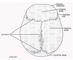
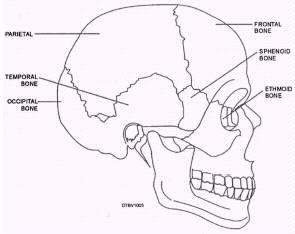
Figure 3-1.\Structure of a typical flat bone. brain, eyes, and ears. It can be divided into two parts: the cranium and the bones of the face. The cranium is primarily involved in housing and protecting the brain. The bones of the face are a complex framework that helps to form facial features, the upper jaw (maxilla) and lower jaw (mandible). With the exception of the mandible and the bones of the inner ear, all skull bones are joined together firmly along seams called sutures. An example of sutures is shown in figure 3-2. Sutures are sometimes considered immovable; however, they do permit a small amount of movement and provide mechanical protection for the brain by absorbing much of the force if a blow to the head occurs. The cranium is formed by eight cranial bones, which form the foundation for attachment of many of the muscles necessary for head movements and chewing. Figure 3-3 show the cranial bones, and Table 3-1 lists them as either single or paired bones. Frontal Bone The frontal bone forms the front part of the skull above the eyes, which includes the forehead and part of
Figure 3-2.\Sutures of a skull.
Table 3-1.\Bones of the Cranium
the nasal cavity. In children, the frontal bone develops as two parts. They are usually fused together by age 5 or 6. The two frontal sinuses (air spaces in the bone) are located above each eye socket. Parietal Bones The two parietal bones are located behind the frontal bone. These bones form the greater part of the right and left sides and the roof of the skull. They each have four borders and are shaped like a curved plate. Temporal Bones The temporal bones form the sides and part of the base of the skull in the area of the ear. One temporal bone is located on each side of the head. It is readily recognized as "fan-shaped." Each encloses the internal ear structures and have depressions called glenoid fossae that forms the articulation with the mandible.
Figure 3-3.\Cranial bones. The zygomatic process of the temporal bone projects out into the zygomatic bone of the face and forms the lateral part of the zygomatic arch. Both the glenoid fossae and zygomatic process can be seen in figure 3-4. |
||
|
||