EXTERNAL ACCESSORY ORGANS
Many of the external accessory organs of the
female reproductive system are referred to collectively
as the vulva. The vulva includes the
labia majora, the labia minora, the
clitoris, and the vestibular glands
(fig. 1-63). The mammary glands are also considered
an accessory organ of the female
reproductive system.
Labia Majora
The function of the labia majora is to enclose and
protect the other external reproductive
organs. The labia majora are composed
of two round folds of fat tissue and a
thin layer of smooth muscle, covered by
skin. On the outer portion of the labia majora, the skin
has numerous hairs, sweat glands, and
sebaceous glands. The inner portion of
skin is thin and hairless. The labia
majora extend from the mons pubis
anteriorly to the perineum (the region between the
vaginal orifice and the anus). The mons
pubis is the pad of fatty tissue
beneath the skin, which overlies the
symphysis pubis.
Labia Minora
Within the labia majora folds are two smaller
folds, called the labia minora. The labia minora extend
from the clitoris to either side of the
vaginal orifice.
Clitoris
The clitoris is a small projectile at the anterior end
of the vulva between the labia minora. It is
richly endowed with sensory nerves that
are associated with the feeling of pleasure
during sexual stimulation.
Vestibule
The vestibule is the area enclosed by the labia
minora that includes that vaginal and urethral
openings. The vestibule contains a pair of
vestibular glands, more commonly known
as the Bartholin's glands. The
Bartholin's glands lay on each side of the
vaginal opening. The ducts of these glands secrete
fluid that moistens and lubricates the
vestibule.
Mammary Glands
The mammary glands, or breasts, are accessory
organs of the female reproductive system. They
develop during puberty under the influence
of the hormones estrogen and
progesterone. The breasts are
responsible for the secretion of milk (lactation) for the
nourishment of newborn infants.
Structurally, the breasts resemble sweat glands. At
the center is a nipple containing 15 to 20
depressions into which ducts from the
lobes of the gland empty. During
pregnancy, placental estrogen and progesterone
stimulate further development of the mammary glands
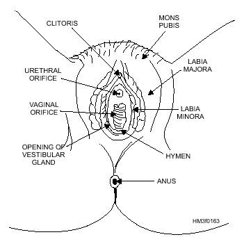
Figure 1-63.-External reproductive organs.
in preparation for lactation. After childbirth, hormones
secreted by the anterior lobe of the
pituitary gland stimulate production
for 6 to 9 months.
|


