Share on Google+Share on FacebookShare on LinkedInShare on TwitterShare on DiggShare on Stumble Upon
Custom Search


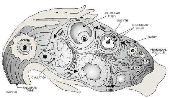
INTERNAL ACCESSORY ORGANS
The internal accessory organs of the female reproductive system include a pair of fallopian tubes, the uterus, and the vagina (fig. 1-61).

Fallopian Tubes
The fallopian tubes, also known as uterine tubes, serve as ducts for the ovaries, providing a passageway to the uterus. The fallopian tubes are composed of three tissue layers. These tissue layers include an inner mucosal layer, a middle muscular layer, and an outer serous layer, and they are continuous with the layers of the uterus. The fallopian tubes are in contact with the ovaries but are not continuous with them. Their funnel-shaped openings, called free openings, are fringed with fingerlike processes that pick up an ovum and draw it into the fallopian tubes. Once the ovum enters the fallopian tubes, it is transported to the uterus by peristalsis and gravity. Fertilization of an ovum normally takes place in the fallopian tubes.

Uterus
The function of the uterus is to receive the embryo that results from the fertilization of an egg cell, and to sustain its life during development. The uterus, or womb, is a hollow, pear-shaped organ with thick, muscular walls. The uterus is divided into two main regions, the body and cervix (fig. 1-61). The body of the uterus consists of the upper two-thirds of the uterus. The cervix is the lower one-third portion of the uterus that projects into the upper part of the vagina. The cervical opening into the vagina is called the external os.

The uterine wall is composed of three layers: the endometrium, the myometrium, and the perimetrium. The inner lining consists of specialized epithelium, called endometrium, which undergoes partial destruction approximately every 28 days in the nonpregnant female. The middle layer, the myometrium, consists of bundles of interlaced muscular fibers. The muscular layer produces powerful rhythmic contractions that are important in the expulsion of the fetus at birth. The perimetrium consists of an outer serosal layer that covers the body of the uterus and part of the cervix. The uterus also has three openings: superiorly and laterally, two openings connect the fallopian tubes to the uterus, and inferiorly, an opening leading to the vagina.

Figure 1-62.-Ovulation process.

Vagina
The vagina is the organ that receives the male sperm during intercourse. It also forms the lower portion of the birth canal, stretching widely during delivery. In addition, it serves as an excretory duct for uterine secretions and menstrual flow.

The vagina is a fibromuscular tube capable of great distention. The canal is approximately 9 cm long and extends from the uterus to the outside. The vaginal orifice is partially closed by a thin membrane of tissue called the hymen. The wall of the vagina consists of three layers. The inner mucosal layer does not have mucous glands; the mucous found in the vagina comes from the glands of the cervix. The middle muscular layer consists mainly of smooth muscles fibers. At the lower end of the vagina is a thin band of smooth muscle that helps close the vaginal opening. The outer fibrous layer consists of dense fibrous connective tissue interlaced with elastic fibers. These fibers attach the vagina to the surrounding organs.


Western Governors University


Privacy Statement - Copyright Information. - Contact Us

Integrated Publishing, Inc. - A (SDVOSB) Service Disabled Veteran Owned Small Business