Custom Search
|
|
 
|
||
|
THE INTEGUMENTARY SYSTEM
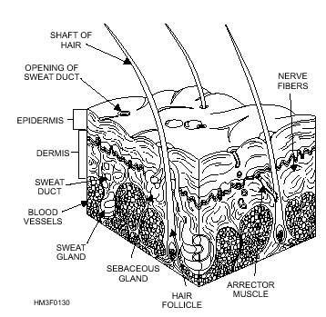
LEARNING OBJECTIVE: Identify skin, its functions, structure, and appendages. Organ systems are comprised of tissues grouped together to form organs, and groups of organs with specialized functions. Since the skin acts with hair follicles, sebaceous glands, and sweat glands, these organs together constitute the integumentary system. SKIN FUNCTION SKIN STRUCTURE Epidermis Dermis BLOOD VESSELS.-The blood vessels of the dermis can dilate to contain a significant portion of the body's blood supply (fig. 1-30). This ability, along with the actions of the sweat glands, forms the body's primary temperature-regulating mechanism. The constriction or dilation of these blood vessels also affects blood pressure and the volume of blood available to the internal organs. NERVE FIBERS.-The skin contains two types of nerve fibers that carry impulses to and from the central nervous system (fig. 1-30). The nerve fibers are distributed to the smooth muscles in the walls of the arteries in the dermis and to the smooth muscles around the sweat glands and hair roots. The first type of nerve fiber carries impulses to the dermal muscles and glands, while the other type carries impulses from sensory receptors (i.e., detecting touch). Both nerve fibers send messages about the external environment to the brain. SMOOTH MUSCLES.-Smooth involuntary muscles are found in the dermis. They are responsible for controlling the skin surface area. When dilated, these muscles allow for maximum skin surface exposure to aid heat loss. When constricted, the skin surface exposure is decreased, thus impeding heat radiation. Repeated muscle contractions (shivering) are also a rapid means of generating body heat. Skin Appendages NAILS.-The nails are composed of horny epidermal scales and are found on the dorsal surfaces of the fingers and toes. They protect the many sensitive nerve endings at the ends of these digits. New formation of nail will occur in the epithelium of the nail bed. As a new nail is formed, the whole nail moves forward, becoming longer. HAIR.-Hair is an epithelial structure found on almost every part of the surface of the body (fig. 1-30). Its color depends on the type of melanin present. The hair has two components: the root below the surface and the shaft projecting above the skin. The root is embedded in a pit-like depression called the hair follicle. Hair grows as a result of the division of the cells of the root. A small muscle, known as the arrector (fig. 1-30), fastens to the side of the follicle and is responsible for the gooseflesh appearance of the skin as a reaction to cold or fear. Each hair follicle is associated with two or more sebaceous glands. SEBACEOUS GLANDS.-Sebaceous glands are found in most parts of the skin except in the soles of the feet and the palms of the hand (fig. 1-30). Their ducts open most frequently into the hair follicles and secrete an oily substance that lubricates the skin and hair, keeping them soft and pliable and preventing bacterial invasion. SWEAT GLANDS.-Sweat glands are found in almost every part of the skin (fig. 1-30). They are control mechanisms to reduce the body's heat by evaporating water from its surface. The perspiration secreted is a combination of water, salts, amino acids, and urea. Normally, about one liter of this fluid is excreted daily. However, the amount varies with atmospheric temperature and humidity and the amount of exercise taken. When the outside temperature is high, or upon exercise, the glands secrete large amounts to cool the body through evaporation. When
Figure 1-30.-Cross section of the skin. evaporation does not remove all the sweat that has been excreted, the sweat collects in beads on the surface of the skin. CERUMINOUS GLANDS.-Ceruminous glands are modified sweat glands found only in the auditory canal. They secrete a yellow, waxy substance called cerumen that protects the eardrum. |
||
|
||