Triceps Brachii
The triceps brachii is the primary extensor of the
forearm (the antagonist of the biceps brachii) (fig. 1-29). It originates at
two points on the humerus and one on the
scapula. These three heads join to form the large muscle
on the posterior surface of the upper arm.
The point of insertion is the olecranon
process of the ulna.
Latissimus Dorsi
The latissimus dorsi is a broad, flat muscle that
covers approximately one-third of the back on each
side (figs. 1-28 and 1-29). It rotates the
arm inward and draws the arm down and
back. It originates from the upper
thoracic vertebrae to the sacrum and the
posterior portion of the crest of the ilium. Its fibers
converge to form a flat tendon that has its
insertion in the humerus.
Gluteus
The gluteus (maximus, minimus (not shown), and
medius) are the large muscles of the
buttocks, which extend and laterally
rotate the thigh, as well as abduct and
medially rotate it (fig. 1-29). They arise from the
ilium, the posterior surface of the lower sacrum, and
the side of the coccyx. Their points of
insertion include the greater
trochanter and the gluteal tuberosity of the
femur. The gluteus maximus is the site of choice for
intramuscular injections.
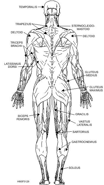
Figure 1-29.-Posterior view of superficial skeletal muscles.
Quadriceps
The quadriceps is a group of four muscles that
make up the anterior portion of the thigh. The four
muscles of this group are the rectus
femoris that originates at the
ilium; and the vastus lateralis, v.
medialis, v. intermedius (not shown), that originate
along the femur (fig. 1-28). All four are
inserted into the tuberosity of the
tibia through a tendon passing over the
knee joint. The quadriceps serves as a strong
extensor of the leg at the knee and flexes the thigh.
Additionally located in the quadriceps area
is the adductor longus that
adducts, rotates, and flexes the thigh.
Biceps Femoris
The biceps femoris (often called the hamstring
muscle) originates at the tuberosity of the ischium (the
lowest portion of the coxal bone, part of
the pelvic girdle) and the middle third
of the femur (fig. 1-29). It is
inserted on the head of the fibula and the lateral
condyle of the tibia. It acts, along with other related
muscles, to flex the leg at the knee and to
extend the thigh at the hip joint.
Gracilis
The gracilis is a long slender muscle located on the
inner aspect of the thigh (figs. 1-28 and
1-29). It adducts the thigh, and flexes
and medially rotates the leg. Its
origin is in the symphysis pubis, and its
insertion is in the medial surface of the tibia, below the
condyle.
Sartorius
The sartorius is the longest muscle in the body. It
extends diagonally across the front of the
thigh from its origin at the ilium,
down to its insertion near the
tuberosity of the tibia (fig. 1-29). Its function is to flex
the thigh and rotate it laterally, and to
flex the leg and rotate it slightly
medially.
Gastrocnemius and Soleus
The gastrocnemius and soleus (together
commonly called the calf muscles) extend the foot at
the ankle (figs. 1-28 and 1-29). The gastrocnemius
originates at two points on the femur; the
soleus originates at the head of the
fibula and the medial border of the
tibia. Both are inserted in a common
tendon called the calcaneus, or Achilles tendon.
Tibialis Anterior
The tibialis anterior originates at the upper half of
the tibia and inserts at the first
metatarsal and cuneiform bones (fig.
1-28). It flexes the foot.
Diaphragm
The diaphragm (not shown) is an internal (as
opposed to superficial) muscle that forms the floor of
the thoracic cavity and the ceiling of the
abdominal cavity. It is the primary
muscle of respiration, modifying the
size of the thorax and abdomen
vertically. It has three openings for the passage of
nerves and blood vessels.
|


