Custom Search
|
|
 
|
||
|
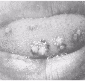
VIRAL INFECTIONS The viral infections of main concern that will be explained are those caused by the herpes simplex virus (HSV), and the human immunodeficiency virus, also referred to as the HIV (causing AIDS) virus. Both are extremely contagious to you and your other dental patients through cross contamination of dental instruments and dental equipment. Also the virus can gain access via the skin, the eye, or mucous membranes. If you treat a patient with one of these or other viruses, ensure you follow the proper infection control procedures outlined in chapter 10. Herpes Simplex Viruses The herpes simplex viruses are among the most common infectious agents. There are two types: Herpes simplex virus\Type 1 (HSV-1) Herpes simplex virus\Type 2 (HSV-2) (genital herpes) In oral pathology the most commonly diagnosed sites for HSV-1 are the oral cavity, tongue (fig. 5-12), lips, and the eyes. Direct contact with HSV-1 lesions is probably the most common mode of spread. Transmission through saliva is possible even if there are no active lesions. Infection on the hands of healthcare personnel from patients shedding HSV can result in herpetic lesions. Other lesions of the HSV-1 virus are acute herpetic gingivostomatitis, characterized by red and swollen gingiva. All of the oral mucosa is tender and eating is painful. Vesicles form throughout the mouth and rupture, leaving painful ulcers.
Figure 5-12.\Herpes simplex virus-Type 1 (HSV-1). The most common of all the herpetic HSV-1 lesions is herpes labialis. They frequently involve the lips and adjacent skin at the corners of the mouth. Recurrence usually starts at the same location, starting with a burning, tingling sensation and then forming vesicles that fuse together leaving large lesions. After the vesicles rupture, crusting of the surface occurs. These lesions are known as "fever blisters." The crusted lesions are also referred to as "cold sores," because a common cold sometimes accompanies these HSV-1 lesions. Known causes for the reoccurrence of the HSV-1 lesions are: Sunlight Menstruation Dental treatment (local trauma) Stress or anxiety The recurrent HSV-1 lesions usually take about 7 to 10 days to resolve. Any routine dental treatment is recommended to be rescheduled during the active phase of these lesions because the disease is highly transmissible. AIDS Virus (HIV Infection) The human immunodeficiency virus type 1 (HIV-l) is the main cause of the acquired immunodeficiency syndrome (AIDS). It is a worldwide epidemic. This deadly disease is a direct threat to all dental health professionals and other healthcare workers who are exposed to patients who carry the virus. Healthcare workers can be exposed to the AIDS virus through contaminated body fluids, exposure to blood or blood products, instruments, and equipment. You should also know some of the oral manifestations that infected people may have. Some of them are the initial signs a dentist can use to diagnosis patients who are carriers of the virus, but who have not been tested or diagnosed. Some of the more common oral manifestations of HIV infection are as follows: Candidiasis\(fig. 5-13) a fungal infection of the mouth, usually red or white in color Hairy leukoplakia\(fig. 5-14) a viral infection, whose lesions appear as white, slightly raised, on the tongue Kaposi's sarcoma\(fig. 5-15) cancerous, dark bluish-purple lesions that involve blood vessels Procedures and precautions for protection will be discussed in chapter 10, "Infection Control." ORAL CANCER Forms of oral cancer are found in the oral cavity at any site, but most often in the tongue, floor of the mouth, and the lower lip. The cancer is a neoplasm
Figure 5-13.\Candidiasis.
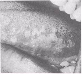
Figure 5-14.\Hairy leukoplakia. (tumor) and is a growth of abnormal tissue. There are two types of neoplasms: Benign tumors\not life threatening Malignant tumors\life threatening if left untreated Classifications of Malignant Tumors Dentists are trained and give special attention when performing an oral cancer screening on a patient to detect any type of cancer. Often these lesions do not cause any pain while in the early stages of development. A malignant tumor can become fatal if not found in its early stages or if left untreated. The following are classifications of malignant tumors. Carcinoma\cancer of the epithelium usually found on the oral mucosa of the mouth, lips (fig. 5-16), tongue, cheeks, and floor of the mouth. Carcinomas start off looking like elevated or ulcerated lesions, and
Figure 5-15.\Kaposi's sarcoma.
Figure 5-16.\Carcinoma of the upper lip. can quickly spread to other locations on the body and invade the lymph nodes. Adenocarcinoma -usually found in the oral region or salivary glands, most often of the palate (fig. 5-17) and appears as a lump or a bulge under the mucosa. Sarcomas-affects the supportive and connective tissues, for example, bones of the jaw. Causes The causes for many neoplasms are unknown. What is known is that the disease is characterized by
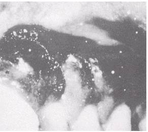
Figure 5-17.\Adenocarcinoma. the abnormal growth and spread of cancer cells. This growth or spread of malignant tumors from one area to another is called metastasis. Modern research concerning the development of neoplasms has been linked to the following factors: Hereditary Chemicals (carcinogens, such as found in tobacco smoke and alcoholic beverages) Overexposure to X-rays Excessive sunlight Smokeless Tobacco Smokeless tobacco, such as chewing tobacco or snuff, may play a role in the development of oral precancerous lesions on the oral mucosa and can result in increased tooth loss from periodontal disease. The area where the user of smokeless tobacco places it in his mouth may leave a smooth or scaly white patch called leukoplakia or snuff-dipper's keratosis (fig. 5-18). Irritation of the oral mucosa occurs because 90 percent of the nicotine of smokeless tobacco is directly absorbed through the oral mucosa, which then goes directly into the blood stream. The effects and damage of nicotine pose a serious health hazard. Many smoking cessation programs are available through naval hospitals and clinics. Dental patients who wish to get assistance from this addiction can be referred to these programs.
Figure 5-18.\Snuff-dipper's keratosis. |
 |
|
|
||