Custom Search
|
|
 
|
||
|
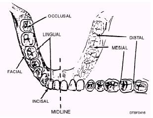
UNIVERSAL NUMBERING SYSTEM The Universal Numbering System is a simplified method of identifying teeth that is approved by the American Dental Association and used by the armed services. This method employs numbers with each tooth designated by a separate number from 1 to 32. Figure 4-15 illustrates the numbering system used on a Standard Dental Chart. When charting, you would refer to a tooth by number rather than the name. Instead of referring to the right maxillary third molar, you would refer to tooth No. 1. Each permanent tooth has its own number. The 20 primary teeth are identified on the dental chart by the use of capital letters A to T. Lettering starts with upper right second primary molar (tooth A, located above the root of the maxillary second premolar); goes across to the upper left second primary molar (tooth J); down to the lower left second primary molar (tooth K), and across to the lower right second primary molar. Please note that the letters of the primary second and first molars appear above the roots of the permanent teeth of the second and first premolars. When using a dental form, remember that the right and left sides are reversed. The right side of the patient's mouth appears on the left side of the dental chart; the left side of the patient's mouth appears on the right side. This arrangement is necessary because the dental officer and the assistant see the sides reversed when they look into a patient's mouth. Full instructions for using dental forms, will be provided in Dental Technician, Volume 2, NAVEDTRA 12573, chapter 2, "Oral Examination." SURFACES OF THE TEETH Not only must the assistant be able to name and locate a tooth, but must also be able to identify the different types of tooth surfaces. Figure 4-16 shows a number of different surfaces of the teeth. Facial, Mesial, Distal, Lingual, and Incisal Surfaces The facial is the surface of a tooth that "faces" toward the lips or cheeks. When there is a requirement to be more specific, terms like labial and buccal are used. The labial is the surface of an anterior tooth that faces toward the lips. The buccal is the surface of a posterior tooth that faces toward the cheek. The mesial is the proximal surface closest to the midline of the arch. The distal is the opposite of mesial. The distal is the proximal surface oriented away from the midline of the arch. The lingual is the surface of an anterior or posterior tooth that faces toward the tongue. Incisal edges are narrow cutting edges found only in the anterior teeth (incisors). Incisors have one incisal edge.
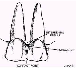
Figure 4-16.\Surfaces of the teeth. Proximal Surfaces A tooth has two proximal surfaces (fig 4-17), one that is oriented toward the midline of the dental arch (mesial) and another that is oriented away from the midline of the arch (distal). Other important surfaces of the proximal area are discussed in the following paragraphs. CONTACT POINT.\The point on the proximal surface where two adjacent teeth actually touch each other is called a contact point. An example of a contact point is when you pass dental floss in between two teeth. You should feel some resistance of the contact point while the floss is being passed through. INTERPROXIMAL SPACE.\The interproximal space is the area between the teeth. Part of the interproximal space is occupied by the interdental papilla. The interdental papilla is a triangular fold of gingival tissue. The part of the interproximal space not occupied is called the embrasure. EMBRASURE. \The embrasure occupies an area bordered by interdental papilla, the proximal surfaces of the two adjacent teeth, and the contact point (fig 4-18). If there is no contact point between the
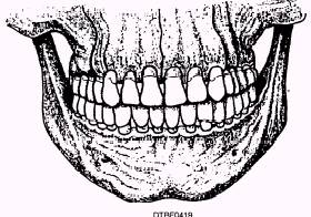
Figure 4-17.\Proximal tooth surfaces and spaces. teeth, then the area between them is called a diastema instead of an embrasure. Occlusal The occlusal surface is the broad chewing surface found on posterior teeth (bicuspids and molars). To get a clearer picture of the various tooth surfaces, refer to figure 4-15, which has previously been discussed. The Dental Chart shows each of the teeth "unfolded" so that the facial, occlusal, incisal, or lingual surfaces of the teeth can be shown. For posterior teeth, the facial surfaces are shown adjacent to the roots, followed by the occlusal surfaces, and then by the lingual surfaces (which are located next to the numbers on the chart). For the anterior teeth, the facial surfaces are shown as a line between the facial and lingual surfaces. The lingual surfaces are located next to the numbers on the chart. OCCLUSION.\Occlusion is the relationship between the occlusal surfaces of maxillary and mandibular teeth when they are in contact. Many patterns of tooth contact are possible. Part of the reason for the variety is the mandibular condyle's substantial range of movement within the temporal mandibular joint. Malocclusion occurs when any abnormality in occlusal relationships exist in the dentition. Centric occlusion, figure 4-19, is the centered contact position of the chewing surfaces of mandibular teeth on the chewing surface (occlusal) of the maxillary teeth. OCCLUSAL PLANE.\Maxillary and mandibular teeth come into centric occlusion and meet along anteroposterior and lateral curves. The anteroposterior curve is called the Curve of Spee
Figure 4-18.\Embrasure.
Figure 4-19.\Centric occlusion. (fig. 4-20) in which the mandibular arch forms a concave (a bowl-like upward curve). The lateral curve is called the Curve of Wilson (fig. 4-21). The composite (combination) of these curves form a line called the occlusal plane, and is created by the contact of the upper and lower teeth as shown in figure 4-22. |
 |
|
|
||