Custom Search
|
|
 
|
||
|
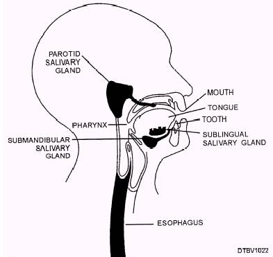
MYLOHYOID AND GENIOHYOID The mylohyoid muscles anatomically and functionally form the floor of the mouth (fig. 3-20). They elevate the tongue and depress the mandible. Their origin is the mandible and insertion is the upper border of the hyoid bone. The geniohyoid muscles are found next to each other, on each side of the midline, directly on top of the mylohyoid muscle. They have the same origin and function as the mylohyoid muscle. PALATE The palate (fig. 3-21) forms the roof of the mouth and is divided into two sections: Hard palate\This section is formed by the palatine process of the maxillary bones and is located in the anterior portion of the roof of the mouth. It has irregular ridges or folds behind the central incisors called rugae. Soft palate\This section forms a soft muscular arch in the posterior part of the palate. The uvula is located on the back portion of the soft palate. When you swallow, the uvula is drawn upward and backward by the muscles of the soft palate. This process blocks the opening between the nasal cavity and pharynx, not allowing food to enter the nasal cavity. The soft palate must function properly to allow good speech quality. Located in the posterior part of the mouth, on both sides of the tongue, are two masses of lymphatic tissue called the palatine tonsils. They assist the body to protect against infections. TEETH The teeth are located in the alveolar process of the maxillae and the mandible. They serve important functions of tearing and masticating food, assisting in swallowing, speaking, and in appearance. The health of the teeth affects the health of the entire body. SALIVARY GLANDS The functions of the three major salivary glands are to keep the lining of our mouths moist, and to bond with food particles creating a lubricant effect that assists in the swallowing process of food. It acts as a cleaning agent to wash away food particles that accumulate in the mouth and on the teeth. Figure 3-22 illustrates the salivary glands. The salivary glands produce two to three pints of saliva daily, which greatly aids in the digestion process. Enzymes are present in saliva, they act on food, and start the breakdown process. In dentistry, knowing exactly where the saliva glands and ducts (openings) are located is important in keeping the mouth dry
Figure 3-20.\Mylohyoid and geniohyoid muscles.
Figure 3-21.\Anatomy of the palate.
Figure 3-22.\Salivary glands. during certain dental procedures. Table 3-5 lists the three major salivary glands. |
||
|
||