Custom Search
|
|
 
|
||
|
THE URINARY SYSTEM
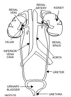
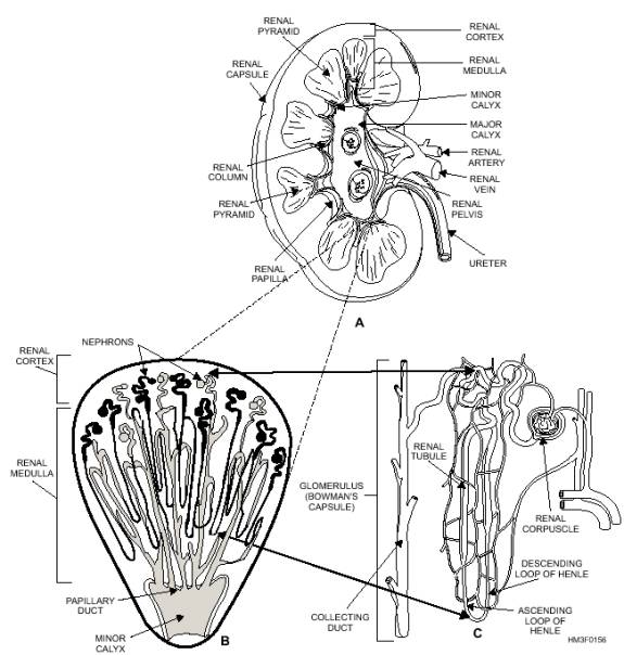
LEARNING OBJECTIVE: Recall the parts of the urinary system and their function(s). The urinary system is the primary filtering system of the body (fig. 1-55). This system is composed of two main organs, the kidneys and urinary bladder. The kidneys produce urine, which is drained from the kidneys by two tubes called ureters. Urine flows down both ureters to the bladder. The urinary bladder is a large reservoir where the urine is temporarily stored before excretion from the body. A tube called the urethra carries the urine from the bladder to the outside of the body. All these parts, except the length of the urethra, are the same in both sexes. KIDNEYS The kidneys are two large, bean-shaped organs designed to filter waste materials from the blood (figs. 1-55 and 1-56). They also assist in controlling the rate of red blood cell formation, and in the regulation of blood pressure, the absorption of calcium ions, and the volume, composition, and pH of body fluids. The kidneys are located in the upper posterior part of the abdominal cavity, one on each side of the spinal column. The upper end of each kidney reaches above the level of the 12th rib. The suprarenal (adrenal) gland sits like a cap on top of each kidney. The kidneys are protected by a considerable amount of fat and supported by connective tissue and the peritoneum. Attached to the hollow side of each kidney is the dilated upper end of the ureter, forming the renal pelvis. Structure The superior end of the ureter forms a funnel-shaped sac called the renal pelvis (fig. 1-56). The renal pelvis is divided into two or three tubes, called major calyces. The major calyces (sing. calyx) are further subdivided into minor calyces. There are groups of elevated projections in the walls of the renal pelvis. These projections are called renal papillae. The renal papillae connect to the minor calyces, through tiny openings in the minor calyces. The principal portion of the kidney is divided into two distinct regions: an inner medulla and outer cortex (fig. 1-56). The renal medulla is composed of pyramid-shaped masses of tubes and tubules called renal pyramids. Renal pyramids drain the urine to the renal pelvis. The renal cortex forms a shell over the renal medulla. Renal cortex tissue dips down, like fingers, between the renal pyramids, and forms what are called renal columns. The cortex possesses very small tubes associated with nephrons. Nephrons are the functional units of the kidneys. RENALBLOOD VESSELS.-The renal artery supplies blood to the kidneys (fig. 1-56). The renal artery enters the kidneys through the hilum, and sends off branches to the renal pyramids. These arterial branches are called interlobar arteries. At the border between the medulla and cortex, the interlobar arteries branch to form the arciform arteries. The arciform arteries branch also and form the interlobular arteries.
Figure 1-55.-The urinary system. NEPHRONS.-The functional units of the kidneys are called nephrons. There are about 1 million nephrons in each kidney. Each nephron consists of a renal corpuscle and a renal tubule (fig. 1-56, view C). The renal corpuscle (Malpighian corpuscle) is composed of a tangled cluster of blood capillaries called a glomerulus. The glomerulus is surrounded by a sac-like structure referred to as the glomerulus yes"> capsule or Bowman's capsule (figs. 1-56, view C, and 1-57). Leading away from the glomerulus is the renal tubule. The initial portion of the renal tubule is coiled and called the proximal convoluted (meaning coiled or twisted) tubule. The proximal convoluted tubule dips down to become the descending loop of Henle. The tubule then curves upward toward the renal corpuscle and forms the ascending loop of Henle. Once the ascending limb reaches the region of the renal corpuscle, it called the distal convoluted tubule. Several distal convoluted tubules merge in the renal Figure 1-56.-Principal parts of the kidney: A. Longitudinal section of a
kidney; B. A renal pyramid containing nephrons; C. A
single nephron. Function Urine is formed through a series of processes in the nephron. These processes are filtration, reabsorption, and secretion. |
||
|
||