Custom Search
|
|
 
|
||
|
DIFFERENTIAL WHITE BLOOD CELL
COUNT
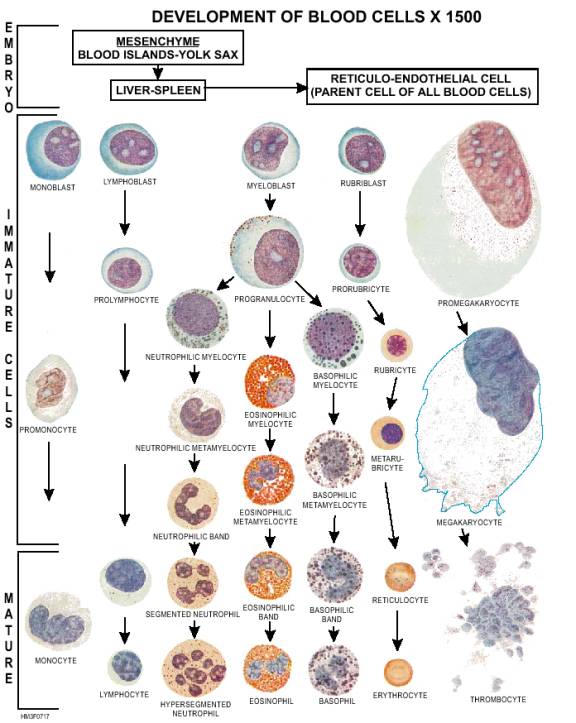
A total white blood cell count is not necessarily indicative of the severity of a disease, since some serious ailments may show a low white cell count. For this reason, a differential white cell count is performed. A differential white cell count consists of an examination of blood to determine the presence and the number of different types of white blood cells. This study often provides helpful information in determining the severity and extent of an infection, more than any other single procedure used in the examination of the blood. The role of white blood cells, or leukocytes, is to control various disease conditions. Although these cells do most of their work outside the circulatory system, they use the blood for transportation to sites of infection. Five types of white cells are normally found in the circulating blood. They are eosinophils, Cell Identification To acquaint you with the developmental stages of each type of leukocyte, a colorized illustration (fig. 7-17) has been provided. This illustration also displays the developmental stages of the red blood cell (erythrocyte) and the blood platelet cell (thrombocyte). To further assist you, identifying characteristics of each type of leukocyte as they appear on a stained blood smear will be covered in the following sections. Laboratories use a blood smear to obtain a differential white cell count. To prepare a blood smear, a blood specimen is spread across a glass slide, stained to enhance leukocyte identification, and examined microscopically. Material requirements and the step-by-step procedure for performing a blood smear will be covered later in this chapter. NEUTROPHILS.-Neutrophils account for the largest percentage of leukocytes found in a normal blood sample, and function by ingesting invading bacteria. On a stained blood smear, the cytoplasm of a neutrophil has numerous fine, barely visible lilac-colored granules and a dark purple or reddish purple nucleus (see figure 7-17). The nucleus may be oval, horseshoe, or "S"-shaped, or segmented (lobulated). Neutrophils are subclassified according to their age or maturity, which is indicated by changes in the nucleus. The subclassifications for neutrophilic cells are metamyelocyte, band, segmented, and hypersegmented. Neutrophilic Metamyelocyte.-A neutrophilic metamyelocyte, also called a "juvenile" cell, is the youngest neutrophil generally reported. The nucleus is fat, indented, and is usually "bean"-shaped or "cashew nut"-shaped (fig. 7-17). Neutrophilic Band.-A neutrophilic band, sometimes called a "stab" cell, is an older or intermediate neutrophil. The nucleus has started to elongate and has curved itself into a horseshoe or S-shape. As the band ages, it matures into a segmented neutrophil (fig. 7-17). Segmented Neutrophil.-A segmented neutrophil is a mature neutrophil. The nucleus of a segmented neutrophil is separated into two, three, four, or five segments or lobes (fig. 7-17). EOSINOPHIL.-Eosinophils aid in detoxification. They also break down and remove protein material. The cytoplasm of an eosinophil contains numerous coarse, reddish-orange granules, which are lighter colored than the nucleus (fig. 7-17). BASOPHIL.-The function of basophilic cells is unknown. It is believed, however, that basophilic cells keep the blood from clotting in inflamed tissue. Scattered large, dark-blue granules that are darker than the nucleus, characterize the cell as a basophil (fig. 7-17). Granules may overlay the nucleus as well as the cytoplasm. LYMPHOCYTE.-The function of lymphocytes is also unknown, but it is believed that they produce antibodies and destroy the toxic products of protein metabolism. The cytoplasm of a lymphocyte is clear sky blue, scanty, with few unevenly distributed, azurophilic granules with a halo around them (fig. 7-17). The nucleus is generally round, oval, or slightly indented, and the chromatin (a network of fibers within the nucleus) is lumpy and condensed at the periphery. MONOCYTE.-The monocyte, the largest of the normal white blood cells, destroys bacteria, foreign particles, and protozoa. Its color resembles that of a lymphocyte, but its cytoplasm is a muddy gray-blue (fig. 7-17). The nucleus is lobulated, deeply indented or horseshoe-shaped, and has a relatively fine chromatin structure. Occasionally, the cytoplasm is more abundant than in the lymphocyte. |
||
|
||