Custom Search
|
|
 
|
||
|
HEMATOCRIT (PACKED CELL VOLUME) DETERMINATION
The hematocrit or packed cell volume (PCV) determines the percentage of red blood cells (RBCs) in whole blood. The normal hematocrit value for men is 42% to 52%; for women, 37% to 47%; and for newborns, 53% to 65%. When hematocrit determinations are below normal, medical conditions such as anemia and leukemia may be present. Above-normal hematocrit determinations indicate medical conditions like dehydration, such as occur in severe burn cases. Currently, automated hematology analyzers supply most hematocrits. However, when hematology analyzers are not available, hematocrit determinations can be manually performed by the microhematocrit method or macrohematocrit method. Both methods call for the blood to be centrifuged, and the percentage of packed red cells is found by calculation. The microhematocrit method is the most accurate manual method of determining blood volume and should be used whenever feasible. Material requirements and the step-by-step procedures for performing the microhematocrit method will be covered in the following sections. Materials Required for Microhematocrit Procedure To perform a hematocrit using the micro- hematocrit method, the following materials are required. Capillary tubes, plain or heparinized 1. Fill the capillary tube two-thirds to three-quarters full with well-mixed, oxalated venous blood or fingertip blood. (For fingertip blood use heparinized tubes, and invert several times to mix.) 2. Seal one end of the tube with clay. 4. Centrifuge at a preset speed of 10,000 to 12,000 rpm for 5 minutes. If the hematocrit exceeds 50 percent, centrifuge for an additional 3 minutes. 5. Place the tube in the microhematocrit reader. Read the hematocrit by following the manufacturer's instructions on the microhematocrit reading device. TOTAL WHITE BLOOD CELL COUNT White blood cell counts are performed either manually or with automated hematology analyzers. Only the manual method will be covered in this chapter. After a brief discussion on abnormal white blood cell counts, we will cover the Unopette method for manually counting white blood cells. Abnormal White Cell Counts
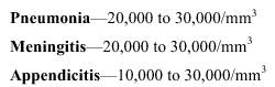
Dyscrasia (the diseased condition) of blood-forming tissues, such as occurs in leukemia (due to a malfunctioning of lymph and marrow tissues) also results in leukocytosis, with extremely high white cell counts. These white cell counts sometimes exceed 1,000,000/mm 3 . Other physiological conditions that can cause leukocytosis and a white cell count as high as 15,000/mm 3 may occur as follows: Shortly after birth Severe or advanced bacterial infections (such as typhoid, paratyphoid, and sometimes tularemia), or when the bacterial infection has been undetected for a period of time (as with chronic beta streptococcal infections of the throat). Infections caused by viruses and rickettsiae, such as measles, rubella, smallpox (until the 4th day), infectious hepatitis, psittacosis, dengue, tsutsugamushi fever, and influenza (when it may fall to 1,500/mm 3 , or shift to leukocytosis if complications develop). Protozoal infections (such as malaria) and helminthic infections (such as trichinosis). (For example, with victims of malaria, slight leukocytosis may develop for a short time during paroxysm (the sudden intensification of symptoms). Shortly thereafter, however, leukopenia ensues.) Overwhelming infections when the body's defense mechanisms break down. Anaphylactic shock |
 
|
|
|
||