

Custom Search
|
|
 
|
||
|
Appendicular Skeleton The appendicular skeleton consists of the bones of the upper and lower extremities. UPPER EXTREMITY.-The upper extremity consists of the bones of the shoulder, the arm, the forearm, the wrist, and the hand (figs. 1-21 and 1-22). The bones that form the framework for the upper extremities are listed in table 1-1. Clavicle.-The clavicle (commonly called the collar bone) lies nearly horizontally above the first rib and is shaped like a flat letter S. The clavicle is a thin brace bone that fractures easily. Its inner end is round and attached to the sternum; its outer end is flattened and fixed to the scapula. The clavicle forms the anterior portion of the pectoral girdle (fig. 1-21). The pectoral girdle is composed of the two clavicles and two scapulae (shoulder blades). It functions as a support for the arms and serves as an attachment for several muscles. Scapula.-The scapula is a triangular bone that lies in the upper part of the back on both sides, between the second and seventh ribs, forming the posterior portion of the pectoral girdle. Its lateral corner forms part of the shoulder joint, articulating with the humerus. Humerus.-The humerus is the longest bone of the upper extremity and is often called the arm bone (fig. 1-22). It articulates with the pectoral girdle to form the shoulder joint, and with the bones of the
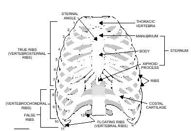
Figure 1-20. 3/4 Anterior view of thorax,
Figure 1-21.-Pectoral girdle.
Table 1-1.-Bones of the Upper Extremities forearm to form the elbow. Its anatomical portions include a head (a rounded portion that fits into a recess of the scapula) called the glenoid fossa; the shaft, which is the main part of the humerus; and the distal end, which includes the prominence (called an epicondyle) and the surfaces that articulate with the bones of the forearm. Radius and Ulna.-When the arm is in the anatomical position with the palm turned forward, the radius is on the lateral (thumb) side and the ulna is on the medial (little finger) side of the forearm (fig. 1-22). When the hand is pronated (with the palm turned downward), the bones rotate on each other and cross in the middle. This pronation makes it possible to turn the wrist and hand (as when opening doors). The ulna and the radius articulate at their proximal ends with the humerus, at their distal ends with some of the carpal bones, and with each other at both ends. |
||
|
||


 style="mso-spacerun: yes">
