Figure 4-10. - Structures of the gingiva.
portion is approximately 1 to 3 mm wide and forms the soft tissue wall of the gingival sulcus next to the tooth. Other structures of unattached gingiva include:
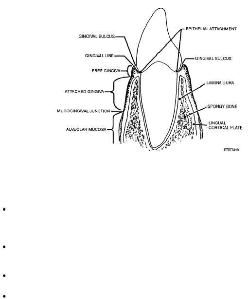
Gingival margin - The 1 mm narrow band of gingiva that forms the immediate collar around the base of the tooth. This area is first to show symptoms of gingivitis.
Gingival sulcus - Area between the unattached gingiva and the tooth. Popcorn hulls get trapped in this area.
Epithelial attachment - Joins the gingiva to the tooth surface.
Interdental papilla - The portion of the free gingiva that fills the interproximal embrasures below the contact areas of adjacent teeth. It helps prevent food from packing between the teeth.
Attached Gingiva. - Located apical to the free gingiva on the labial and lingual aspects.
It is firmly fixed to the underlying bone of the cortical plates of the alveolar process.
Mucogingival Junction.-A line that separates the attached gingiva from the lining mucosa.
Lining Mucosa
Lining mucosa is found on the inside of the lips, cheeks, vestibule, soft palate, and under the tongue. It consists of a thin, fragile tissue that is very vascular. Lining mucosa is brighter red in color than masticatory mucosa. Also included in the lining mucosa is alveolar mucosa. It lies apical to the mucogingival junction and is loosely attached.
TOOTH MORPHOLOGY
This section describes the external features of the teeth. A tooth is defined as "one of the hard, bony appendages that are borne on the jaws...and serve for the seizing and mastication of food, as weapons of offense and defense, etc."
In man and the lower animals, the design of the teeth are a reflection of eating habits.
Animals, classified according to their eating habits, are carnivorous (flesh eating), herbivorous (plant eating), or omnivorous (eating everything; both flesh and plant).
TYPES OF TEETH
Man is omnivorous, so his teeth are formed for cutting, tearing, and grinding food.
The human permanent dentition is divided into four classes of teeth based on appearance and function or position.
Figure 4-11 illustrates the types and working surfaces of the four classes of teeth.
Incisors
Incisors are named because they are used to incise food. They are located in the front of the mouth and have sharp, thin edges for cutting. The lingual surface can have a shovel-shaped appearanc Continue Reading