cancellous bone. When viewed by a radiograph, trabecular bone has a web-like appearance.
Alveolar Bone Proper
The alveolar bone proper is a thin layer of compact bone, that is a specialized continuation of the cortical plate and forms the tooth socket. The lamina dura (fig. 4-10) is a horseshoe shape white line on a dental radiograph that roughly corresponds to the alveolar bone proper.
PERIODONTAL LIGAMENT
The periodontal ligament (fig. 4-8) is a thin, fibrous ligament that connects the tooth to the bony socket. Normally, teeth do not contact the bone directly; a tooth is suspended in its socket by the fibers of the ligament. This arrangement allows each tooth limited individual movement. The fibers act as shock absorbers to cushion the force of the chewing impact of mastication.
TISSUES OF THE ORAL CAVITY
The oral cavity is made up of specialized epithelial tissues that surround the teeth and serve as a lining. These tissues are called the oral mucosa and consist of three types: masticatory mucosa, lining mucosa, and specialized mucosa.
Masticatory Mucosa
Masticatory mucosa is comprised of the tissue that covers the hard palate and the gingiva.
Masticatory mucosa is usually light pink in color (can vary with skin color) and is keratinized. Keratinized tissue has a horny, tough, protective outer layer of tissue. Characteristics of masticatory mucosa are:
no submucosa lies under the masticatory mucosa,
held in place firmly to bone and does not move,
has a dense, hard covering, and
functions to withstand the active process of chewing and swallowing food.
HARD PALATE.
- The hard palate or roof of the mouth (fig. 4-9) is covered with masticatory mucosa and is firmly adhered to the palatine process (bone). Its 4-7
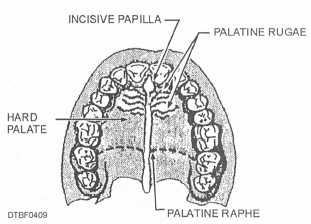
Figure 4-9. - Masticatory mucosa of the hard palate.
color is usually pale pink. Important structures of the hard palate are:
Incisive papilla - Located at the midline, directly posterior of the maxillary central incisors (pear-shaped in appearance).
Palatine raphe - Extends from the incisive papilla posteriorly at the midline (may be ridge shaped in appearance with a whitish streak at the midline).
Palatine rugae - Extends laterally (along side) from the incisive papilla and from the palatine raphe (wrinkled, irregular ridges in appearance).
GINGIVA. - The gingiva, shown in figure 4-10, is specialized masticatory mucosa covering the alveolar process. In a healthy mouth, gingiva is firmly in place encircling the necks of the teeth. It aids in the support of the teeth, and protects the alveolar process and periodontal ligament from bacterial invasion. Healthy gingiva is firm and resilient. Healthy gingiva under normal flossing and brushing activities does not bleed. The color of healthy gingiva can range from pale pink to darker shades (purple to black) depending on each individual’s pigmentation. The surface of the attached gingiva and interdental papillae may be stippled (resembling the texture of the skin of an orange).
Like the tongue, the gingiva is highly vascular and receives its blood supply from the lingual, mental, buccal, and palatine arteries. Other important aspects of the gingiva are discussed in the following paragraphs.
Unattached Gingiva. - The portion of gingiva that extends from the gingival crest to the crest of the bone is called unattached gingiva. It can also be called the free gingiva. It can be displaced and is not bound directly to the tooth or bone. In a healthy mouth, this
Continue Reading