Custom Search
|
|
 
|
||
|
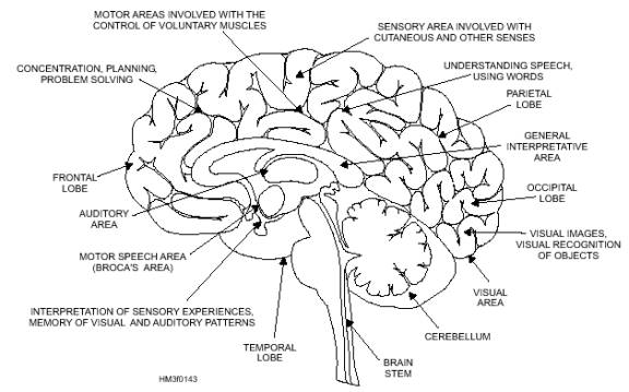
Spinal Cord The spinal cord is continuous with the medulla oblongata and extends from the foramen magnum, through the atlas, to the lower border of the first lumbar vertebra, where it tapers to a point. The spinal cord is surrounded by the bony walls of the vertebral canal (fig. 1-44). Ensheathed in the three protective meninges and surrounded by fatty tissue and blood vessels, the cord does not completely fill the vertebral canal, nor does it extend the full length of it. The nerve roots serving the lumbar and sacral regions must pass some distance down the canal before making their exit. The sympathetic trunk contains the paravertebral ganglia (sing. ganglion), knotlike masses of nerve cell bodies (fig. 1-44). Across section of the spinal cord shows white and gray matter (fig. 1-45). The outer white matter is composed of bundles of myelinated nerve fibers arranged in functionally specialized tracts. It establishes motor communication between the brain and the body parts. The inner gray unmyelinated Figure 1-43.-Functional areas of the brain.
Figure 1-44.-Spinal cord. The spinal cord may be thought of as an electric cable containing many wires (nerves) that connect parts of the body with each other and with the brain. Sensations received by a sensory nerve are brought to the spinal cord, and the impulse is transferred either to the brain or to a motor nerve. The majority of impulses go to the brain for action. However, a system exists for quickly handling emergency situations. It is called the reflex arc. If you touch a hot stove, you must remove your hand from the heat source immediately or the skin will burn very quickly. But the passage of a sense impulse to the brain and back again to a motor nerve takes too much time. The reflex arc responds instantaneously to emergency situations like the one just described. The sensation of heat travels to the spinal cord on a sensory nerve. When the sensation reaches the spinal cord, it is picked up by an interneuron in the gray matter. This reception then triggers the appropriate nerve to stimulate a muscle reflex drawing the hand away. An illustrated example of the reflex arc is shown in figure 1-45. The reflex arc works well in simple situations requiring no action of the brain. Consider, however, what action is involved if the individual touching the stove pulls back and, in so doing, loses balance and has to grab a chair to regain stability. Then the entire spinal cord is involved. Additional impulses must travel to the brain, then down to the muscles of the legs and arms to enable the individual to maintain balance and to hold on to a steadying object. While all this activity is going on, the stimulus is relayed through the sympathetic autonomic nerve fibers to the adrenal glands, causing adrenalin to flow, which stimulates heart action. The stimulus then moves to the brain, making the individual conscious of pain. In this example, the spinal cord has functioned not only as a center for spinal relaxes, but also as a conduction pathway for other areas of the spinal cord to the autonomic nervous system and to the brain. |
 
|
|
|
||