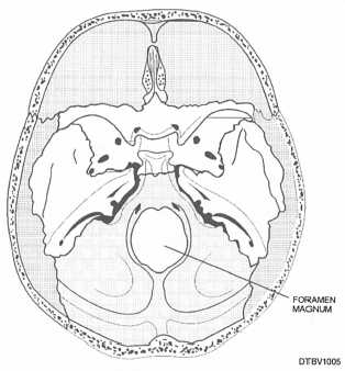
Figure 3-5. - Foramen magnum of cranial cavity viewed from above.
The occipital bone is an irregular, four-sided bone that is somewhat curved upon itself.
Sphenoid Bone
The sphenoid bone has a wing-like shape and is internally wedged between several other bones in the front part of the cranium (fig. 3-6). This bone assists with the formation of the base of the cranium, the sides of the skull, and the floors and sides of the orbits.

Figure 3-6. - Sphenoid bone viewed from above.
Ethmoid Bone
The ethmoid bone is situated in front of the sphenoid bone in the front part of the cranium (fig. 3-7). Through small openings in this bone pass nerves to the roof of the mouth that are responsible for sense of smell.
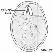
Figure 3-7. - Ethmoid bone viewed from above.
BONES OF THE FACE
The facial skeleton consists of 14 stationary bones and a mobile lower jawbone (mandible). These 14 bones (table 3-2) form the basic shape of the face, and are responsible for providing attachments for muscles that make the jaw move and control facial expressions. Figures 3-8 and 3-9 show the bones of the face.
Maxillae Bones
The maxillae bones are the largest bones of the face and together form the upper jaw. The maxilla (singular) consists of a body and. four processes: zygomatic, frontal, alveolar and palatine. The maxilla forms the hard palate, floor of the nose, part of the orbits (eye sockets), and the tooth sockets of the upper teeth. Above the roots of the upper teeth and below the
Table 3-2. - Bones of the Face
| Single Bones | Paired Bones |
| Vomer | Maxillary |
| Mandible | Palatine |
| Zygomatic | |
| Lacrimal | |
| Nasal | |
| Inferior nasal conchae |