well as in the duct glands and in the skin. Under a microscope, the smooth muscle fiber lacks the striped appearance of other muscle tissue (fig. 3-25). This tissue is also called INVOLUNTARY muscle because it is not under conscious control.
The CARDIAC MUSCLE tissue forms the bulk of the walls and septa (partitions) of the heart, as well as the origins of the great blood vessels. The fibers of the cardiac muscle differ from those of the skeletal and smooth muscles in that they are shorter and branch into a com- plicated network (fig. 3-26). The cardiac muscle has the most abundant blood supply of any mus- cle in the body, receiving twice the blood flow of the highly vascular skeletal muscles and far more than the smooth muscles. Cardiac muscles con- tract to pump blood out of the heart and through the cardiovascular system. Interference with the blood supply to the heart can result in a heart attack.
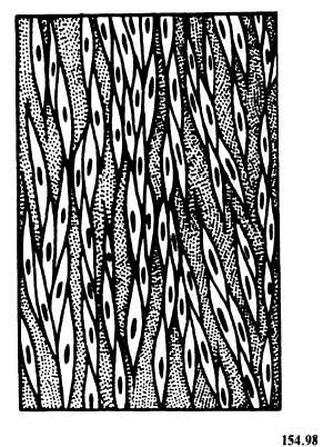
Figure 3-25.—Smooth muscle fibers.
IMPORTANT FUNCTIONAL MUSCLES
These muscles, shown in figures 3-27 and 3-28, are described below.
The MASSETER muscle raises the mandible, or lower jaw, to close the mouth. It is the chewing muscle in the mastication of food. It originates in the zygomatic process and adjacent parts of the maxilla and is inserted in the mandible.
The TEMPORAL muscle assists the masseter and draws the mandible backward. It has its origin in the temporal fossa and is inserted in the cor- onoid process of the mandible.
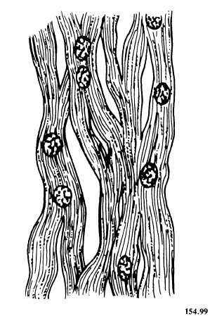
Figure 3-26.—Cardiac muscle fibers.
The STERNOCLEIDOMASTOID muscles are located on both sides of the neck. Acting in- dividually, these muscles rotate the head left or right. Acting together, they bend the head for- ward toward the chest. The sternocleidomastoid muscle originates in the sternum and clavicle and is inserted in the mastoid process of the temporal bone. This muscle is commonly affected in cases of stiff neck.
The TRAPEZIUS muscles are a broad, trapezium-shaped pair of muscles on the upper back, which raise or lower the shoulders. They cover approximately one-third of the back. They originate in a large area, which includes the 12 thoracic vertebrae, the seventh cervical vertebra,